Now you can visualize proteins, RNA, RNA-protein complex and other biomolecules using ResourceFunction BioMoleculePlot3D in Wolfram Language. Source - https://resources.wolframcloud.com/FunctionRepository/resources/BioMoleculePlot3D/
Here are some examples of what this function can do:
Here are some examples of what this function can do:
Draw structures of proteins using four-letter PDB ID
Draw structures of proteins using four-letter PDB ID
In[]:=
ResourceFunction["BioMoleculePlot3D"]["1crn"]
Out[]=
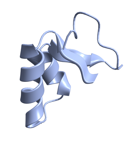
Define colors of individual chains in the biomolecule
Define colors of individual chains in the biomolecule
In[]:=
ResourceFunction["BioMoleculePlot3D"]["2H7Z","ChainStyle"->{RGBColor[0.70135,0.093019,0.00140383],RGBColor[0.289647,0.222614,0.484169]}]
Out[]=
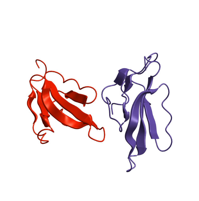
Visualize individual residues in a biomolecule along its backbone
Visualize individual residues in a biomolecule along its backbone
In[]:=
ResourceFunction["BioMoleculePlot3D"]["7KAG",PlotLegends->Automatic,"ColorScheme"->"Residue"]
Out[]=
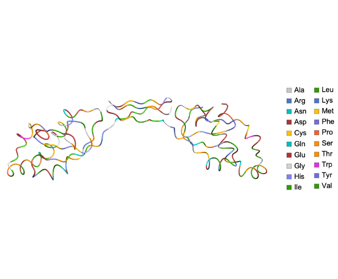
Visualize RNA-protein complex
Visualize RNA-protein complex
In[]:=
ResourceFunction["BioMoleculePlot3D"]["1I6U","ChainStyle"->{Hue[3/20],Hue[0.7],Hue[0.8],Hue[1]}]
Out[]=
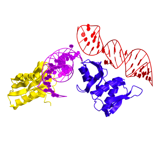
Visualize protein ligand complex
Visualize protein ligand complex
Some cool protein structures
Some cool protein structures
Structure of green fluorescent protein from the jellyfish Aequorea victoria, which has a β - barrel motif :
Acknowledgements
Acknowledgements
I would like to acknowledge the co-authors of the function Jan Mangaldan and Jason Biggs for their contributions, suggestions and guidance.