The present study investigates how explicit solvation affects the interaction between D-glucuronic acid (D-GlcA), a major component of the Cryptococcus neoformans capsule, and metal cations such as Ca²⁺. Using HF and DFT calculations, the binding of D-GlcA to fully solvated Ca(H₂O)₆²⁺ complexes was modeled to explore how water coordination alters reaction energies and geometry. Geometry optimizations and energy comparisons were used to evaluate how hydration influences metal–ligand bond lengths, electron redistribution, and complex stability. The influence of cation radius and charge on complex formation was explored by substituting Ca²⁺ with Y³⁺ to assess how these properties affect structural distortion and thermodynamic favorability. While calcium and yttrium followed similar overall bonding trends, yttrium complexes exhibited greater magnitudes of structural fluctuation and a highly endothermic second binding step. These findings would enhance the mechanistic model for metal-mediated capsule clumping in C. neoformans, with implications for understanding fungal virulence and informing antifungal strategies targeting ion-mediated aggregation.
Introduction
Introduction
Cryptococcus neoformans is a pathogenic yeast responsible for approximately 200,000 deaths each year worldwide, with the majority occurring in Sub-Saharan Africa. One of its most critical virulence factors is its polysaccharide capsule, which enables the yeast to evade host immune defenses and modulate the immune response, thereby enhancing its survival and pathogenicity.
Glucuronic Acid
Glucuronic Acid
Glucuronic acid with the carboxylic acid group highlighted.
In[]:=
MoleculePlot3D[Molecule["(2S,3S,4S,5R,6R)-3,4,5,6-Tetrahydroxyoxane-2-carboxylic acid"],MoleculePattern[{"H","O","C","O"},{Bond[{1,2},"Single"],Bond[{2,3},"Single"],Bond[{3,4},"Double"]}]]
Out[]=

The polysaccharide capsule of C. Neoformans is primarily composed of complex polysaccharides, including mannose, xylose, and glucuronic acid (GlcA). These sugar units are arranged in a highly branched, negatively charged matrix that plays a vital role in immune evasion and structural integrity. Glucuronic acid, in particular, introduces carboxylic acid functional groups into the capsule structure, contributing to its overall negative charge and providing potential binding sites for divalent and trivalent metal cations. These interactions are hypothesized to influence capsule organization and cell aggregation.
Aim
Aim
This investigation examines how variations in metal cation charge and ionic radius affect their interaction energies with deprotonated glucuronic acid (D-GlcA), a key component of the Cryptococcus neoformans capsule. Prior evidence suggests that calcium-mediated crosslinking between GlcA residues may contribute to capsule aggregation. To more accurately represent physiological conditions, this study models Ca²⁺ as a metal hydrate via the Ca(H₂O)₆²⁺ complex and explores how explicit hydration impacts metal–ligand energetics. Substituting calcium with other cations further allows for comparison across ionic sizes and charges.
Understanding these interactions has implications for fungal pathogenicity and may inform future therapeutic strategies aimed at disrupting capsule integrity or aggregation associated with cryptococcal meningitis.
Understanding these interactions has implications for fungal pathogenicity and may inform future therapeutic strategies aimed at disrupting capsule integrity or aggregation associated with cryptococcal meningitis.
Schrödinger Equation and Approximation
Schrödinger Equation and Approximation
This study employs the time-independent Schrödinger equation as the basis for electronic structure calculations, which are essential for optimizing molecular geometries and evaluating interaction energies.
𝐻 Ψ = 𝐸 Ψ
The Hamiltonian operator 𝐻 represents the total energy of a molecular system—including kinetic energy of the electrons, electron-nuclear attraction, and electron-electron repulsion.
The wave function Ψ contains all information about the system’s quantum state, and 𝐸 is the corresponding energy eigenvalue. Solving this equation would, in theory, yield a complete description of a molecule’s electronic structure. However, for systems with more than one electron, exact solutions cannot be expressed in terms of elementary functions, as the integrals—especially those accounting for electron repulsion—typically do not have analytical, closed-form solutions.
To address this, approximation methods such as Hartree-Fock (HF) are employed. Hartree-Fock approximates the many-electron wavefunction by representing electrons as moving independently within an average potential generated by all other electrons. This approach yields a single-determinant wavefunction that serves as a practical starting point for modeling molecular interactions. In this study, HF forms the initial basis for modeling interactions between D-glucuronic acid and solvated metal dications.
Optimizing Calcium Complexes using Hartree-Fock
Optimizing Calcium Complexes using Hartree-Fock
To construct the hexaaqua calcium complex—a neutral metal hydrate—the function was used. Each atom was manually placed, and bonds were defined accordingly to reflect the correct coordination geometry.
Molecule
Load the quantum chemistry paclet & build the calcium hexaaqua species:
In[]:=
Needs["WolframChemistry`QuantumChemistry`"]
In[]:=
mol=Molecule;
Building and Optimizing Molecular Structures
Building and Optimizing Molecular Structures
The function was used to set the formal charge of the calcium ion to +2, making the overall structure positively charged:
MoleculeModify
In[]:=
molCaDication=MoleculeModify[mol,{"SetFormalCharge",{1->+2}}]
Out[]=
Molecule


Starting structure of the Ca(H₂O)₆²⁺ complex:
In[]:=
MoleculePlot3D[molCaDication]
Out[]=

Since the and functions define only basic bonding relationships and do not account for electronic interactions, the resulting structure is unlikely to represent the lowest-energy geometry.
Molecule
MoleculeModify
To optimize the molecular structure of the Ca(H₂O)₆²⁺ complex, the Hartree-Fock method with the STO-3G basis set was used as the default approximation:
In[]:=
default=ModelChemistry["HartreeFock","sto-3g"]
Out[]=
ModelChemistry[HartreeFock,sto-3g]
Using the default parameter defined above, the Ca(H₂O)₆²⁺ dication was optimized and imported via :
CloudGet
In[]:=
optCaDication=CloudGet
Out[]=
ElectronicStructureResult

Optimized structure of the Ca(H₂O)₆²⁺ complex:
In[]:=
MoleculePlot3D[optCaDication["Molecule"],AtomLabels->"AtomIndex"]
Out[]=

Compute the symmetry of the optimized structure:
In[]:=
optCaDication["Molecule"]["PointGroupDisplay"]
Out[]=
D
3
The Ca(H₂O)₆²⁺ complex shown above exhibits D₃ symmetry, characterized by two key symmetry elements: a C₃ principal rotation axis that passes through the central calcium ion and remains symmetrical when rotated 60 degrees, and three perpendicular C₂ axes that bisect the angles between the adjacent ligand pairs. This symmetry results in a trigonal prismatic coordination geometry, where the six water molecules are arranged symmetrically around the calcium ion.
The presence of D₃ symmetry reduces the number of unique integrals required during quantum mechanical calculations, thereby lowering computational cost and runtime while preserving accuracy.
The presence of D₃ symmetry reduces the number of unique integrals required during quantum mechanical calculations, thereby lowering computational cost and runtime while preserving accuracy.
Display the symmetry elements in the Ca(H₂O)₆²⁺ complex:
In[]:=
ResourceFunction["MoleculeSymmetryPlot3D"][optCaDication["Molecule"]]
In[]:=
molGlcA=Molecule["(2S,3S,4S,5R,6R)-3,4,5,6-Tetrahydroxyoxane-2-carboxylic acid"]
Out[]=
Molecule

Starting structure of the glucuronic acid molecule:
In[]:=
MoleculePlot3D[molGlcA]
Out[]=

The function was used again for the water and hydronium yielded as products:
Molecule
In[]:=
water=Molecule[{Atom["H"],Atom["O"],Atom["H"]},{Bond[{1,2},"Single"],Bond[{2,3},"Single"]}]
Out[]=
Molecule
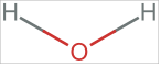
Starting structure of the water molecule:
In[]:=
MoleculePlot3D[water]
Out[]=

Starting structure of the hydronium molecule:
Starting structure of the [Ca(H₂O)₄D-GlcA]⁺ complex:
Optimized structure of the water molecule:
Optimized structure of the hydronium molecule:
Optimized structure of the glucuronic acid molecule:
Optimized structure of the [Ca(H₂O)₄D-GlcA]⁺ complex:
Calculating ΔE
Calculating ΔE
The change in electronic energy (ΔE) represents the difference in heat content between products and reactants under constant pressure conditions. In computational chemistry, ΔE is typically approximated using single-point electronic energies obtained from quantum chemical calculations. Since these calculations do not directly account for vibrational, rotational, or translational contributions, the electronic energy difference is used as an approximation:
Reaction 1 is as follows:
Then, all single point energies for the five molecules were calculated below:
Formula used to calculate ΔE:
Using this information, the net change in energy for reaction 1 was calculated:
Starting structure of the Ca(H₂O)₂(D-GlcA)₂ complex:
The molecular geometry was optimized using the model chemistry defined above.
Optimized structure of the Ca(H₂O)₂(D-GlcA)₂ complex:
Reaction 2 is as follows:
By combining this with previously obtained data, the net energy change for reaction 2 can be obtained:
The net energy change for the overall mechanism was also calculated:
Optimizing Calcium Complexes using Density Functional Theory
Optimizing Calcium Complexes using Density Functional Theory
After initial geometry optimizations using the Hartree-Fock method with the STO-3G basis set, further refinement was carried out using Density Functional Theory (DFT). Because Hartree-Fock neglects dynamic correlation—specifically the Coulomb repulsion between electrons—DFT with Local Density Approximation (LDA) was employed to provide a more accurate treatment of electron correlation through exchange-correlation functionals.
Instead of using on the default settings from above, the dft parameter was defined to perform Density Functional Theory calculations. The STO-3G basis set was preserved due to its broad element compatibility.
Newly Optimizing Previous Molecules using DFT
Newly Optimizing Previous Molecules using DFT
All of the previous molecules were newly optimized:
To maintain consistency in models and basis sets throughout the mechanism, the H₂O, H₃O⁺, and GlcA molecules were also geometry-optimized using the same DFT model chemistry.
Optimized structure of the Ca(H₂O)₂(D-GlcA)₂ complex using DFT:
Calculating ΔE
Calculating ΔE
The single-point energy was then calculated for the newly optimized molecular structures:
Using these values, the energy change of each mechanism was calculated as follows:
Analysis
Analysis
The energy changes (ΔE) associated with each reaction step in the proposed mechanism were calculated using both Hartree-Fock (HF) and Density Functional Theory (DFT) methods. The HF calculations yielded a total ΔE of 1010.72 kJ/mol (390.781 kJ/mol + 619.937 kJ/mol), while the DFT calculations produced a total ΔE of 1392.78 kJ/mol (315.149 kJ/mol + 1077.63 kJ/mol). Both methods resulted in positive ΔE values, indicating that each step and the overall two-step reaction were endothermic, requiring an input of energy to proceed.
The overall energy required to bind two D-glucuronic acid (D-GlcA) molecules to the metal center was therefore 1010.72 kJ/mol according to the HF method and 1392.78 kJ/mol according to the DFT method.
The overall energy required to bind two D-glucuronic acid (D-GlcA) molecules to the metal center was therefore 1010.72 kJ/mol according to the HF method and 1392.78 kJ/mol according to the DFT method.
The discrepancy between these values can be attributed to the fundamental differences between the two computational approaches. Hartree-Fock theory treats electrons within a mean-field approximation, neglecting electron correlation effects, which generally leads to an underestimation of the total energy. In contrast, DFT incorporates electron correlation through exchange-correlation functionals, typically providing a more accurate approximation of the single-point energies of each molecule. Consequently, the higher ΔE obtained via DFT reflects its improved treatment of electron interactions and is considered more reliable for describing the energetics of the system. For this reason, the previously used value for the energy change was rejected in favor of the DFT results.
Alternative Metal Cation(s)
Alternative Metal Cation(s)
The objective was to determine whether replacing the central calcium cation with other metal cations would affect the binding. The investigation tested whether both the charges and the ionic radii of the cations had any impact on the net energy change and the metal ligand bonding. This was accomplished by selecting appropriate metal cations with a coordination number of VI after consulting the Database of Ionic Radii and comparing the results to the original calcium complexes and reaction energies. These calculation were done using DFT (LDA + VWN) and the STO-3G basis set.
Yttrium
Yttrium
The Yttrium(III) ion (Y³⁺) with a coordination number of VI has a crystal radius of 1.04Å and ionic radius of 0.9Å, which falls right within the acceptable range of ±0.1Å.
Then, the MoleculeDraw function was used to add D-glucuronic acid in place of water ligands on the equatorial planes:
Each of the Yttrium-D-GlcA complexes was then optimized using the DFT on the basis set of “STO-3G”:
Optimized structure of the Y(H₂O)₆³⁺ complex:
Optimized structure of the [Y(H₂O)₄D-GlcA]²⁺ complex:
Optimized structure of the [Y(H₂O)₂(D-GlcA)₂]⁺ complex:
The Single Point energies of the DFT-optimized Yttrium-glucuronic acid complexes were calculated:
Reaction 3 is as follows:
Reaction 4 is as follows:
Analysis
Analysis
The calculated energy changes for the yttrium mechanism revealed a distinctive energetic profile. The first reaction step was exothermic, with a ΔE of–120.59 kJ/mol, indicating an initial release of energy upon binding D - glucuronic acid (D - GlcA) to the metal center. In contrast, the second step was highly endothermic, requiring an energy input of approximately + 1.21*10 ⁶ kJ/mol. Consequently, the overall reaction was strongly endothermic, with a total ΔE of + 1.21*10 ⁶ kJ/mol. This large energy requirement suggests a significant energetic barrier in the second step, which will be further examined in the following section on the structural comparison of metal cation complexes.
Structural Comparison of Metal Cation Complexes
Structural Comparison of Metal Cation Complexes
In order to investigate the mechanisms underlying C. Neoformans cell aggregation, changes in metal–oxygen bonding were analyzed as the system transitioned from metal–aqua complexes to metal–aqua–D-glucuronic acid (D-GlcA) complexes. To evaluate potential electron redistribution associated with metal binding, particular attention was given to the bond lengths involving oxygen atoms—specifically the Metal–O, Metal–O–C, Metal–O=C, and C=O (carbonyl group) motifs. Variations in these bond lengths served as indicators of electron density shifts to or from the metal center. The following tables and analyses summarized these observations.
Calcium
Calcium
Ca-O Bond
Ca-O Bond
The mean calcium–oxygen bond length measured between the calcium ion and coordinated water molecules was calculated:
Mean Ca-O bond length in the Ca(H₂O)₆²⁺ complex:
Mean Ca-O bond length in the [Ca(H₂O)₄D-GlcA]⁺ complex:
Individual Ca-O bond lengths in the [Ca(H₂O)₄D-GlcA]⁺ complex:
The overall Ca–O bond lengths increased by approximately 0.1 Å—from 2.296 Å to 2.409 Å—upon the addition of the first D - GlcA molecule to the initial Ca (H₂O) ₆²⁺ complex, with two notably elongated bonds measuring 2.348 Å and 2.460 Å, respectively.
Mean Ca-O bond length in the Ca(H₂O)₂(D-GlcA)₂ complex:
The addition of a second D-GlcA molecule resulted in slightly strengthened Ca–O interactions, as indicated by a modest decrease in average bond length to 2.397 Å.
Ca-O-C Bond
Ca-O-C Bond
The mean calcium–oxygen bond length involving the deprotonated hydroxyl oxygen of D-GlcA was obtained below:
Ca-O-C bond length in the [Ca(H₂O)₄D-GlcA]⁺ complex:
Mean Ca-O-C bond length in the Ca(H₂O)₂(D-GlcA)₂ complex:
The Ca–O–C bond lengths in the Ca(H₂O)₂GlcA₂ complex both decreased—though to different extents—indicating a more stable interaction between the calcium dication and each coordinating oxygen atom compared to those observed in the [Ca(H₂O)₄GlcA]⁺ complex.
Ca-O=C Bond
Ca-O=C Bond
The mean calcium–oxygen bond length involving the double-bonded (carbonyl) oxygen of D-GlcA was calculated:
Ca-O=C bond length in the [Ca(H₂O)₄D-GlcA]⁺ complex:
Mean Ca-O=C bond length in the Ca(H₂O)₂(D-GlcA)₂ complex:
The metal–oxygen bond was weakened, as indicated by an overall increase in bond length of approximately 0.09 Å, suggesting a reduction in electron density around the calcium dication.
Carbonyl Bond
Carbonyl Bond
A key aspect of this reaction was the carbonyl bond, as the oxygen atom possesses a lone pair of electrons. As previously noted, the Ca–O=C bond length decreased, which indicates that electron density from the lone pair was transferred to strengthen the C=O bond.
In order to verify the above claim, the mean carbonyl bond length was calculated for each calcium complex:
Carbonyl bond length in GlcA prior to coordination with the metal center:
Carbonyl bond length in the [Ca(H₂O)₄D-GlcA]⁺ complex:
Mean carbonyl bond length in the Ca(H₂O)₂(D-GlcA)₂ complex:
It was observed that the bond length decreased each time D-GlcA was added to the complex, which suggests that in both mechanisms, the carbonyl acted as the electron density acceptor.
Yttrium
Yttrium
The metal-oxygen bond lengths for Yttrium complexes were also tabulated in the same manner:
Y-O Bond
Y-O Bond
The mean yttrium–oxygen bond length measured between the calcium ion and coordinated water molecules was calculated:
Mean Y-O bond length in the Y(H₂O)₆³⁺ complex:
Mean Y-O bond length in the [Y(H₂O)₄D-GlcA]²⁺ complex:
Individual Y-O bond lengths in the [Y(H₂O)₄D-GlcA]²⁺ complex:
Similar to what was observed from the Ca-O bond, the overall Y-O bond lengths saw an increase by approximately 0.04Å—from 1.997Å to 2.04Å—upon the addition of the first D-GlcA molecule to the initial Y(H₂O) ₆³⁺ complex, with two notably changed bonds measuring 1.993 Å and 2.117Å, respectively.
Mean Y-O bond length in the [Y(H₂O)₂(D-GlcA)₂]⁺ complex:
The addition of a second D-GlcA molecule resulted in the strongest Y-O bond strength of all three yttrium complexes, which is shown by the shortest mean bond length 1.938Å.
Y-O-C Bond
Y-O-C Bond
The mean yttrium–oxygen bond length involving the deprotonated hydroxyl oxygen of D-GlcA was obtained below:
Y-O-C bond length in the [Y(H₂O)₄D-GlcA]²⁺ complex:
Mean Y-O-C bond length in the [Y(H₂O)₂(D-GlcA)₂]⁺ complex:
Unlike the case from Ca-O-C bond, the Y-O-C bond lengths in [Y(H₂O)₂GlcA₂]⁺ complex increased significantly—with approximately 0.1Å and 0.3Å increase respectively—which demonstrates a remarkable decline in bond strength and stability when another D-GlcA molecule was added to the [Y(H₂O)₄GlcA]²⁺ complex.
Y-O=C Bond
Y-O=C Bond
The mean yttrium–oxygen bond length involving the double-bonded (carbonyl) oxygen of D-GlcA was calculated:
Y-O=C bond length in the [Y(H₂O)₄D-GlcA]²⁺ complex:
Mean Y-O=C bond length in the [Y(H₂O)₂(D-GlcA)₂]⁺ complex:
Similar to the previous result, the metal–oxygen bond experienced a slight weakening as indicated by the increased bond length, suggesting a reduction in electron density around the calcium dication. However, the degree of bond weakening was substantially greater in this case, with the metal-oxygen bond length increasing by 0.6Å compared to a 0.09Å increase observed previously.
Carbonyl Bond
Carbonyl Bond
The mean carbonyl bond length was calculated for each yttrium complex:
Carbonyl bond length in GlcA prior to coordination with the metal center:
Carbonyl bond length in the [Y(H₂O)₄D-GlcA]²⁺ complex:
Mean carbonyl bond length in the [Y(H₂O)₂(D-GlcA)₂]⁺ complex:
Consistent with the results for the calcium complexes, the carbonyl acted as the electron density acceptor, which is proven by the decrease in bond length.
Notable Differences Between Calcium and Yttrium Complexes
Notable Differences Between Calcium and Yttrium Complexes
Differences Observed in Structural Changes
Differences Observed in Structural Changes
Although the overall trend of metal–oxygen bond lengthening upon D-GlcA addition was largely preserved across all metal centers, distinct differences emerged in both the pattern and magnitude of these structural responses. In calcium complexes, the average Ca–O bond length increased modestly from 2.296 Å to 2.409 Å upon the addition of the first D-GlcA ligand, and then decreased slightly to 2.397 Å with the second. These fluctuations were relatively small, indicating that the calcium coordination sphere remained largely stable and resistant to significant geometric distortion.
However, more prominent changes were observed in the Ca–O–C interactions. Upon coordination of a second GlcA ligand, the Ca–O–C bond lengths decreased—though asymmetrically—suggesting the development of tighter and more directional interactions with the GlcA framework. This stabilization appeared to occur at the expense of the Ca–O=C bond, which elongated in the Ca(H₂O)₂(D-GlcA)₂ complex, likely due to electron density redistribution away from the carbonyl site. Such behavior is consistent with calcium' s relatively rigid coordination preferences and its tendency to accommodate additional ligands without inducing major structural reorganization.
In contrast, the yttrium complexes exhibited a structural response that, while more prominent in magnitude, closely mirrored the overall trend observed in the calcium system. The average Y–O bond lengths fluctuated with successive ligand additions: initially increasing from 1.997 Å to 2.040 Å upon coordination of the first D-GlcA ligand, then decreasing to 1.938 Å in the [Y(H₂O)₂GlcA₂]⁺ complex. This pattern mirrors the behavior of the calcium complexes, where a slight elongation was followed by contraction, suggesting that the underlying coordination mechanism remains largely consistent across both metal centers. Despite yttrium’s higher charge and marginally smaller ionic radius, the binding mechanism largely appeared similar with no noticeable difference in structure. However, the magnitude of change was greater in the yttrium complexes, suggesting a more loose coordination structure.
The Y–O–C framework, in particular, demonstrated notable divergence in local bonding behavior. The Y–O=C interactions underwent significant destabilization upon the addition of the second GlcA ligand, with bond lengths increasing by approximately 0.1 Å and 0.3 Å, respectively. This selective elongation suggests that, although the overall Y–O coordination became more compact, certain donor interactions were compromised—likely due to increased steric congestion or less favorable orbital alignment. Nevertheless, the resemblance in structural response between the yttrium and calcium complexes implies that the metal’s formal charge plays a limited role in determining the direction of ligand-induced geometric changes. Instead, the differences appear to stem primarily from the scale of reorganization rather than a fundamental shift in binding mechanism.
Differences in Energy Changes
Differences in Energy Changes
Both the calcium and yttrium mechanisms were overall endothermic, indicating that an input of energy was required to bind two D-glucuronic acid (D-GlcA) molecules to the respective metal centers. However, the yttrium mechanism exhibited an exothermic first step, releasing –120.59 kJ/mol of energy, in contrast to the calcium mechanism where both reaction steps were endothermic. This initial energy release in the yttrium mechanism may be attributed to differences in coordination geometry and the nature of ligand–metal interactions. Specifically, yttrium, as a larger and more electropositive metal cation with a higher coordination number, could form stronger initial interactions with D-GlcA or induce a more stable intermediate complex during the first step, thereby lowering the system’s energy. Such differences in metal ion size, charge density, and electronic structure are known to influence coordination modes and binding affinities, which likely contributed to this divergence in reaction energetics.
The difference in the magnitudes of the energy changes between the two mechanisms was also remarkable. The calcium mechanism proceeded via two moderately endothermic steps, with an overall energy requirement of +1392.78 kJ/mol, indicative of a relatively gradual energy input needed to achieve full binding. However, the yttrium mechanism’s second step required an exceptionally high energy input of approximately +1.21 × 10⁶ kJ/mol, resulting in a substantially larger overall endothermic reaction. This contrast suggests that the second step in the yttrium mechanism may involve a highly unfavorable rearrangement or dissociation process, possibly due to steric hindrance, ligand strain, or disruption of coordination environments that are less problematic in the calcium system. Furthermore, the difference in electronic structure between calcium (a divalent alkaline earth metal) and yttrium (a trivalent transition metal) likely alters the metal–ligand bonding dynamics, including bond strengths and electron density distributions, which can significantly affect reaction thermodynamics.
The unusually high energy demand observed in the yttrium mechanism’s second step suggests either a particularly unfavorable reaction pathway or potential limitations inherent in the computational approach for this system.
The unusually high energy demand observed in the yttrium mechanism’s second step suggests either a particularly unfavorable reaction pathway or potential limitations inherent in the computational approach for this system.
Conclusion
Conclusion
This study computationally investigated the binding interactions between D-glucuronic acid (D-GlcA) and metal dications, focusing on calcium and yttrium, with explicit solvation modeled via hexaaqua metal complexes. Molecular geometries were optimized using Hartree-Fock and Density Functional Theory (DFT) with the STO-3G basis set to assess how solvation affects structural features and relative binding energies. Comparisons across one- and two-ligand complexes revealed trends in bond lengths, coordination geometries, and energy changes. Calcium and yttrium followed similar structural patterns—initial metal–oxygen bond elongation followed by contraction—suggesting a conserved binding mechanism, though yttrium exhibited larger structural shifts and a more extreme energetic profile. These findings refine current models of ion-mediated aggregation in the Cryptococcus neoformans capsule and provide a foundation for future studies under biologically relevant conditions.
Future Directions
Future Directions
Due to constraints in computational resources and time, higher-accuracy methods beyond the Local Density Approximation (LDA) formulation of Density Functional Theory (DFT), as well as metal cations requiring basis sets more sophisticated than STO-3G, could not be employed in the present study. Future work will aim to overcome these constraints by incorporating broader and more accurate basis sets (e.g., split-valence and triple-zeta sets) and by employing higher-level electronic structure methods such as unrestricted DFT, Configuration Interaction (CI), and Coupled Cluster (CC) techniques. These improvements would enable a more complete and quantitatively reliable understanding of metal–ligand interactions, particularly in systems involving heavy or open-shell transition metals.
In addition, future investigations could explore alternative binding sites within the glucuronic acid molecule that may yield lower single-point energies. Identifying such sites could offer deeper insight into the most thermodynamically favorable coordination geometries and further elucidate the role of electron delocalization in stabilizing metal–ligand complexes.
Moreover, it is essential to optimize each complex within a more realistic solvation environment—comprising not only pure water but also physiologically relevant media such as saline solutions—given that these interactions occur in aqueous biological systems rather than in the gas phase or under dry conditions.
References
References
Acknowledgements
Acknowledgements
I would like to thank my mentor, Jason Sonnenberg, for his guidance and unconditional support throughout this project. This research would not have been possible without his patience, steady encouragement, and unwavering support—even through my late-night questions, moments of doubt, and more than a few mental breakdowns. I am also grateful to Gracen Gerbig from the Johns Hopkins Bloomberg School of Public Health for introducing me to an entirely new branch of biochemistry and for allowing me to join her ongoing research. I would like to extend my sincere appreciation to Jason Biggs, who developed the QuantumChemistry paclet and thereby enabled me to perform HF/DFT calculations with ease. I am also thankful to the program directors—Eryn Gillam, Rory Foulger, and Megan Davis—as well as to Stephen Wolfram, for ensuring that my research journey was both smooth and enjoyable. Lastly, I am grateful to the other mentors I consulted throughout this project and to those who offered their moral support along the way.
CITE THIS NOTEBOOK
CITE THIS NOTEBOOK
Analyzing metal interactions with Cryptococcus Neoformans
by Eileen "Hyunseo" Lee
Wolfram Community, STAFF PICKS, July 10, 2025
https://community.wolfram.com/groups/-/m/t/3501894
by Eileen "Hyunseo" Lee
Wolfram Community, STAFF PICKS, July 10, 2025
https://community.wolfram.com/groups/-/m/t/3501894