Introduction
Introduction
Wolfram Language is a multi-paradigm programming language designed for functional programming. It is mainly used in Wolfram Alpha and Mathematica.
This year, Texas Tech University organized a HACKATHON with the purpose of detecting and classifying breast cancer using Wolfram Language. This competition aims to introduce girls from Colegios Científicos to programming, promote female participation in STEAM fields, and support the development of future professionals in a growing country such as Costa Rica.
The competition focuses on participants demonstrating the skills and knowledge acquired during their preparation. They analyze a code provided by the organizers to identify possible errors and correct them appropriately.
This document presents essential information to understand the operation of the code developed by the CCAtlántico team. The document includes the following sections:
- Code functionality
- Input
- Output
- Team conclusions
- Extra challenge
- References
HACKATHON2025


Input
First Section: Directories and Link Paths
First Section: Directories and Link Paths
01: Defining directories
01: Defining directories
Directories are essential when coding. Defining the paths where our files are located is important so the platform can use them correctly without issues. A folder called “CCATLANTICO” was defined as the main directory for the code. Inside this folder, the notebook is stored which allows effective access to the information.
The SetDirectory command is used to define the main directory, while the Directory command confirms that the notebook recognizes the directory. This indicates that everything is correctly set and we can continue. Double backslashes (\\) are used so the command works properly.
The SetDirectory command is used to define the main directory, while the Directory command confirms that the notebook recognizes the directory. This indicates that everything is correctly set and we can continue. Double backslashes (\\) are used so the command works properly.
In[]:=
SetDirectory["D:\\BIRADS"]
Out[]=
D:\BIRADS
In[]:=
Directory[]
Out[]=
D:\BIRADS
02: Mammogram Paths
02: Mammogram Paths
Inside the main folder just created, there were added two additional folders along with the notebook.
Both folders contained a large database of mammograms, which were provided before the challenge by the Texas Tech Hackathon 2025 team.
Dataset 1: CBIS-DDSM: Breast Cancer Image Dataset. 5.3GB Zip: https://www.kaggle.com/datasets/awsaf49/cbis-ddsm-breast-cancer-image-dataset
Dataset 2: Mammogram Mastery: A Robust Dataset for Breast Cancer Detection and Medical Education. 933 MB Zip https://data.mendeley.com/datasets/fvjhtskg93/1
Within the database titled “Mammogram Mastery: A Robust Dataset for Breast Cancer Detection and Medical Education”, there is a folder called “Breast Cancer Dataset”. As its name indicates, this folder contains both augmented images (“Augmented Dataset”) and original images (“Original Dataset”) of the mammograms.
The most important folders are those that contain Cancer and NoCancer images.
Modifications were added, including the BI-RADS classifications, where classifications 1, 2, and 3 were defined as NoCancer, and classifications 4 and 5 were defined as Cancer.
The “Breast Imaging Reporting and Data System” (BI-RADS) classification is a reporting system used to describe the results of mammograms. In a range of zero to six, from normal or benign (non-cancerous) to malignant (cancerous).
BI-RAD 0 “Inconclusive”: The radiologist may have observed a suspicious lesion but it is not clearly defined. An additional mammogram of the area of interest or an additional ultrasound is required to compare the results with previous studies.
BI-RAD 1 “Normal”: No suspicious images were identified, structures are normal and the study is considered negative because there are no abnormal findings. The patient should continue with annual mammography.
BI-RAD 2 “Benign findings (non-cancerous)”: The study shows benign findings and no signs of cancer. The patient should continue with annual mammography.
BI-RAD 3 “Findings probably bening”: Less than 2% risk of malignancy. Findings in this category have a very high probability (greater than 98%) of being non-cancerous. It’s recommended an immediate evaluation with additional projections or ultrasound, and short term follow up every 6 months for two years.
BI-RAD 4 “Suspicious abnormality”: It’s recommended to consider a biopsy to characterize the findings and obtain an accurate diagnosis.
BI-RAD 5 “Abnormality that suggests a malignant finding”: The findings have the appearance of cancer and there is a high probability that it is cancer.
BI-RADS 6 “Already proven malignant lesion”: Mammogram findings that have already been shown to be cancerous. The mammogram is ordered to see how the cancer is responding to therapy.
Each image was reviewed individually by category. The images classified as non-cancer were analyzed, and based on the characteristics of classifications 1, 2 and 3, they were manually separated. The cancer images were separated according to the characteristics and details observed in the images, corresponding to category 4 and above.
The non-cancer images (categories 1 and 2) generally showed uniform tissue patterns, smooth and well-defined borders, and consistent intensity without irregular structures. In contrast, the cancer images (category 3 and above) presented heterogeneous textures, irregular or spiculated margins, asymmetry, and localized regions with abnormal density or brightness that differed clearly from the surrounding tissue.
Based on these characteristics, the original dataset was manually restructured to create a modified dataset classified according to BI-RADS categories 1 to 5. This new dataset was then used to train and evaluate the model, enabling it to distinguish between normal findings, benign lesions, probably benign findings, suspicious abnormalities, and highly suggestive malignant lesions.
https://drive.google.com/file/d/1j7DYyFcc8XY2KeBgXE4hAen8agHRvLEO/view?usp=sharing
03: Defining Mammogram Paths
03: Defining Mammogram Paths
Five link paths were defined: examBIRADS1, examBIRADS2, examBIRADS3, examBIRADS4, and examBIRADS5.
After that, these paths were converted into variables. This allows the use of Wolfram Language commands on the images contained in these folders.
After that, these paths were converted into variables. This allows the use of Wolfram Language commands on the images contained in these folders.
In[]:=
examBIRADS1="D:\\BIRADS\\BIRADS NoCancer\\BIRADS 1";examBIRADS2="D:\\BIRADS\\BIRADS NoCancer\\BIRADS 2";examBIRADS3="D:\\BIRADS\\BIRADS NoCancer\\BIRADS 3";examBIRADS4="D:\\BIRADS\\BIRADS Cancer\\BIRADS 4";examBIRADS5="D:\\BIRADS\\BIRADS Cancer\\BIRADS 5";
04: Variable Manipulation to Confirm Correct Setup
04: Variable Manipulation to Confirm Correct Setup
The birads1Images variable is defined using the FileNames command. This command ensures that whenever this variable is called, it refers to all images (Infinity) in the birads1 folder, regardless of the file type (“*”).
examImagesBIRADS1=FileNames["*",examBIRADS1,Infinity];examImagesBIRADS2=FileNames["*",examBIRADS2,Infinity];examImagesBIRADS3=FileNames["*",examBIRADS3,Infinity];examImagesBIRADS4=FileNames["*",examBIRADS4,Infinity];examImagesBIRADS5=FileNames["*",examBIRADS5,Infinity];
In this part of the code, commands such as:
- Length: are used to verify that the platform is using all the images
In[]:=
Length[examImagesBIRADS1]Length[examImagesBIRADS2]Length[examImagesBIRADS3]Length[examImagesBIRADS4]Length[examImagesBIRADS5]
Out[]=
8002
Out[]=
95
Out[]=
131
Out[]=
412
Out[]=
119
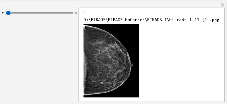
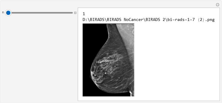
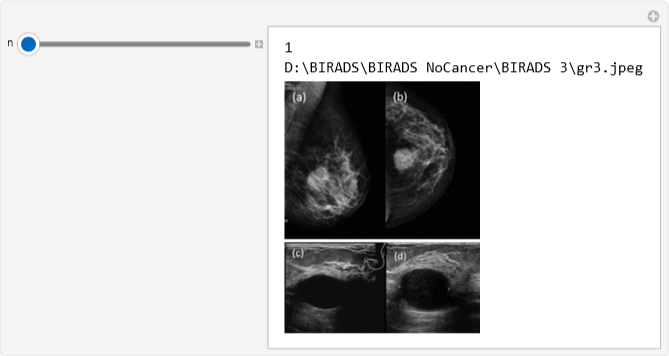
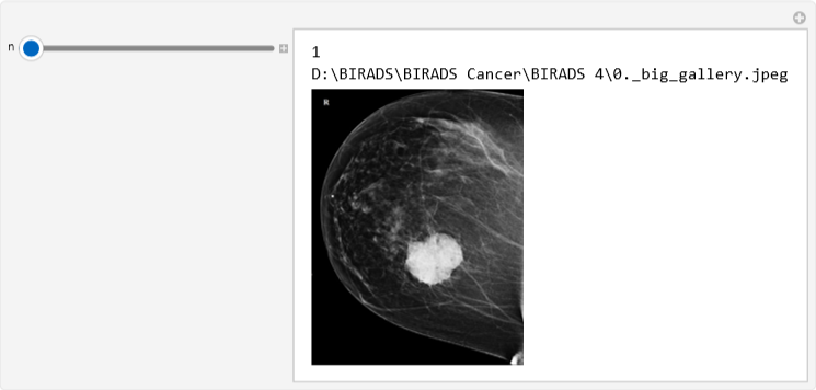
- Manipulate: displays all the images in the folders, allowing them to be viewed directly within the code once
In[]:=
Manipulate[Column[{n,examImagesBIRADS1[[n]],Image[Import[examImagesBIRADS1[[n]]],ImageSize->Small]}],{n,1,Length[examImagesBIRADS1],1},SaveDefinitions->True]
Out[]=
In[]:=
Manipulate[Column[{n,examImagesBIRADS2[[n]],Image[Import[examImagesBIRADS2[[n]]],ImageSize->Small]}],{n,1,Length[examImagesBIRADS2],1},SaveDefinitions->True]
Out[]=
In[]:=
Manipulate[Column[{n,examImagesBIRADS2[[n]],Image[Import[examImagesBIRADS2[[n]]],ImageSize->Small]}],{n,1,Length[examImagesBIRADS2],1},SaveDefinitions->True]
Out[]=
In[]:=
Manipulate[Column[{n,examImagesBIRADS3[[n]],Image[Import[examImagesBIRADS3[[n]]],ImageSize->Small]}],{n,1,Length[examImagesBIRADS3],1},SaveDefinitions->True]
Out[]=
In[]:=
Manipulate[Column[{n,examImagesBIRADS4[[n]],Image[Import[examImagesBIRADS4[[n]]],ImageSize->Small]}],{n,1,Length[examImagesBIRADS4],1},SaveDefinitions->True]
Out[]=
Second Section: AI Training with Databases
Second Section: AI Training with Databases
01: TrainingData and the Beginning of Machine Learning
01: TrainingData and the Beginning of Machine Learning
This section introduces the main concepts related to Wolfram Artificial Intelligence and Machine Learning, which are key elements in the development of this prototype. Machine Learning is a subfield of Artificial Intelligence in which machines are trained to become more autonomous, to predict data, and mainly to learn from the information provided. The AI must be trained using the variables defined previously. However, the images in the folders need to be edited to make recognition easier. For this reason, two variables were created: TrainingCancerData for examCancer and TrainingNoCancerData for examNoCancer.
These variables are defined using several commands:
- ColorConvert: converts the images in the database to grayscale- ImageResize: resizes the images to 600 pixels
- “&/@”: works as the Map command, which is essential to apply a function to variables
- RandomSample: applies this process to a random sample of 100 images. A larger range allows more data to be loaded and can lead to better results from the Artificial Intelligence
02: Importance of Testing at This Stage
02: Importance of Testing at This Stage
The test was performed using the Length command. As in previous cases, this command allows us to view the number of images being used. A result of 100 indicates that the process was completed without errors and that the model successfully learned from the 100 loaded images. Likewise, if more images are added, the Length result would reflect that new quantity.
Third Section: AI Training with Databases
Third Section: AI Training with Databases
01: Functionality of the “Classify” Command
01: Functionality of the “Classify” Command
The purpose of this coding process is to correctly detect cancer in the mammograms that are uploaded. The input allows the user to upload an image, while the output displays the diagnosis. This process is possible thanks to the Classify tool. This command assigns a label to the result of the image recognition. If the image matches the Cancer data from the database, the cancerClassify variable indicates the presence of cancer. The same process applies when cancer is not detected.
This variable is defined using the Classify command.
This variable is defined using the Classify command.
Finally, ClassifierFunction works as a function that indicates the class in which the data is most likely to belong.
02: Testing for this section
02: Testing for this section
Lastly, the Information command is used to obtain details about the data. This information confirms that everything is correctly set and that the system is functioning properly.
03: Function of some commands
03: Function of some commands
- CloudPut[cancerClassify, “cancerClassifier”]: Is used to publish the data to the cloud.
- BarChart: The original code used PieChart; however, a BarChart was chosen because it provides clearer visualization and better organization.
- Information: Displays the information together with a chart.
Fourth Section: Downloading User Data Uploaded to the Cloud
Fourth Section: Downloading User Data Uploaded to the Cloud
01: Function of the “CloudDeploy” command
01: Function of the “CloudDeploy” command
The final part of this code begins with commands that allow the development of the microsite. This specific command makes it possible to deploy the previous code to the cloud.
As shown, this command includes another one called FormFunction, which creates a form interface where the user can upload images. Then, using the CloudGet command, the images are retrieved from the cloud where they were uploaded and later analyzed by the cancerClassifier.
As shown, this command includes another one called FormFunction, which creates a form interface where the user can upload images. Then, using the CloudGet command, the images are retrieved from the cloud where they were uploaded and later analyzed by the cancerClassifier.
Output
Image of the page where both the mammogram and the patient’s data are attached:
Conclusions
Conclusions
◼
I. Use of Artificial Intelligence and the Concept of Machine Learning in Medical Fields
In the field of medicine, specifically in diagnostic medicine, the use of technology is common to achieve accurate disease diagnosis. Mammograms, for example, use a technique known as tomosynthesis, which applies X-rays to create three dimensional images of the breast.
However, despite advances in imaging techniques, the real challenge begins where technology ends: in the interpretation of results and the optimization of the diagnostic workflow. This does not mean replacing humans, on the contrary, it is beneficial for doctors’ health, as it helps reduce fatigue and overload caused by analyzing a large number of cases.
Artificial Intelligence (AI) is an emerging technology that has transformed many areas, especially Oncology. Concepts such as Machine Learning allow AI systems to be trained to identify cancer presence in diagnostic images. This supports automation and improves the efficiency of these processes, which are essential for human health.
◼
II. Importance of the Programming Language: Wolfram Language
Wolfram Language is a programming language well suited for working with large amounts of data. Its ability to perform complex operations using only a few lines of code, together with its built in Artificial Intelligence, allows the creation of stable and highly optimized tools for automation tasks.
◼
III. Personal Conclusions
- Helena: BI-RADS Classification
She was responsible for organizing and classifying the mammogram images according to the BI-RADS scale. This required separating the images into cancer and no-cancer categories and further subdividing them by BI-RADS classification. One of the main challenges was analyzing patterns, densities, and characteristics of the mammograms, which required careful biological analysis.
She was responsible for organizing and classifying the mammogram images according to the BI-RADS scale. This required separating the images into cancer and no-cancer categories and further subdividing them by BI-RADS classification. One of the main challenges was analyzing patterns, densities, and characteristics of the mammograms, which required careful biological analysis.
- Valerie: Model Programming and Language Understanding
She was responsible for the development and training of the Machine Learning model, as well as the organization and structure of the code. She worked with the new database and ensured that all parts of the system functioned correctly. The main difficulty was integrating all parts of the project into a functional system, as well as working with large and heavy datasets.
She was responsible for the development and training of the Machine Learning model, as well as the organization and structure of the code. She worked with the new database and ensured that all parts of the system functioned correctly. The main difficulty was integrating all parts of the project into a functional system, as well as working with large and heavy datasets.
- Vieleny: Written Content and Documentation
She was responsible for the development, structure, and coherence of the written content, including the essay and technical documentation. She translated the programming logic into a storytelling-style explanation, making the system understandable for readers without a technical background. One of the main challenges was explaining the code clearly using simple language, while also managing the overall writing workload of the project.
She was responsible for the development, structure, and coherence of the written content, including the essay and technical documentation. She translated the programming logic into a storytelling-style explanation, making the system understandable for readers without a technical background. One of the main challenges was explaining the code clearly using simple language, while also managing the overall writing workload of the project.
Challenge
Challenge
How could this tool help a clinic in the interior of Costa Rica?
How could this tool help a clinic in the interior of Costa Rica?
Breast cancer is a type of malignant tumor that develops when cells in breast tissue begin to grow uncontrollably. It can affect both men and women, however, approximately 99% of breast cancer cases occur in women, while 0.5% to 1%occur in men. According to the World Health Organization (WHO), in 2022, 2.3 million women were diagnosed with breast cancer worldwide and 670,000 deaths were recorded that same year.
In Costa Rica, the Ministry of Health and the National Institute of Statistics and Census (INEC) reported 398 cases in 2022, as well as 426 deaths among women of all ages due to this disease.
One of the most important factors in overcoming breast cancer, as with many other types of cancer, is early detection. However, the Costa Rican health system faces two major limitations that directly affect early detection: the high concentration of radiologists in the Central Valley and the long waiting times for mammograms to be interpreted by specialists.
When a woman undergoes a mammogram, the image must be reviewed and interpreted by a medical specialist, usually a radiologist. In Costa Rica, as in many countries, there is a shortage of specialists. If an excessive number of breast cancer exams are performed in a year, patients may wait more than five years to receive final results.
Many countries in Europe and Asia already use or are developing Artificial Intelligence systems for breast cancer detection through medical images. This is also possible in Costa Rica. The implementation of a well-trained AI system would allow rural medical centers (in areas such as Talamanca, Upala, or Buenos Aires de Puntarenas) to receive immediate results.
This would help specialists reduce waiting times by allowing them to discard cases where breast cancer is not present and prioritize those where it is suspected.
AI would not replace medical specialists, but instead support them by accelerating the detection process and enabling early treatment, which in breast cancer can make the difference between life and death.
In a country that is territorially small but has unequal distribution of medical resources, such as specialists, Artificial Intelligence could help provide access to specialized diagnosis throughout the country. This would ensure that a woman in Limón or Puntarenas receives the same diagnostic quality as a woman in San José.
Telling our history to the world
Telling our history to the world
In Central America there’s a small country called Costa Rica, with an even smaller town called Limon, the seventh out of seven provinces. A special place mainly because of its location, covering the southern Caribbean coast of the country, and its cultural diversity, with heritage from Afro-descendant, Indigenous, Afro-Caribbean, Chinese, and white cultures.
With a population of 470,383 inhabitants distributed across six cantons, 116,457 of them live in the central canton, where are the main ports of the country. Limon is known for its port activity and agricultural exports, specially bananas, pineapples, cocoa, sugar and coffee.
Important, because Costa Rica has a high dependence in exportation, pillar in the economy, employment and foreign investment. People in this canton are mainly engaged in port, customs, agriculture, tourism, commerce and fishing activities.
Ending last year, the university Texas Tech in Costa Rica and wolfram research organized a challenge known as “hack with purpose”, a hackathon for girls in the National System of Scientific High Schools. Limón brought their own team, called the CCAtlantico, after the high school located in the province.
Three girls, Valerie Artavia, Helena León and Vieleny McCalla came back to their town with the third place after creating a prototype with machine learning that was able to identify cancer presence or absence in mammograms. They also represented data collection to create a “medical record” for each patient and a classification in the BY-RADS scale of mammal cancer.
This wasn’t just an achievement for the Atlantic scientific high school (CCA) or for the team. It was also a opportunity to learn and conscience about breast cancer awareness and the importance of mammograms. In Costa Rica, this type of cancer is the leading cause of death from tumor in women and an early diagnosis increase the possibility of cure by up to 95%.
DataSets
DataSets
To ensure the validity of our project information, we have included links to the specific databases used for the prototype. These databases contain the information used (BIRADS Images 1-5) so that users can view the sources and verify the results obtained.
https://drive.google.com/file/d/1j7DYyFcc8XY2KeBgXE4hAen8agHRvLEO/view?usp=sharing
https://drive.google.com/file/d/1j7DYyFcc8XY2KeBgXE4hAen8agHRvLEO/view?usp=sharing
References
References
During the final stage of the hackathon, which involved developing corrections to the code provided by the organizers, the participants of the CCAtlántico team (Valerie, Helena, and Vieleny) consulted various web pages to ensure that the final work was supported by solid and reliable information in order to achieve the best possible results. These sources are part of the material provided by the organizers.
Thank You!
CITE THIS NOTEBOOK
CITE THIS NOTEBOOK
A computational approach to early breast cancer detection using Wolfram
by Valerie Artavia, Helena León, and Vieleny McCalla
Wolfram Community, STAFF PICKS, March 5, 2026
https://community.wolfram.com/groups/-/m/t/3649858
by Valerie Artavia, Helena León, and Vieleny McCalla
Wolfram Community, STAFF PICKS, March 5, 2026
https://community.wolfram.com/groups/-/m/t/3649858